

The Power of Precision
High Definition Intravascular Imaging
High Definition Intravascular Imaging
High-definition intravascular imaging is a cutting-edge medical technology that provides detailed and real-time visualization of blood vessels from within. It allows medical professionals to observe and diagnose cardiovascular conditions with exceptional clarity and precision. By utilizing advanced imaging techniques, this technology enhances the accuracy of diagnosis and improves treatment outcomes.
Types of High-Definition Intravascular Imaging
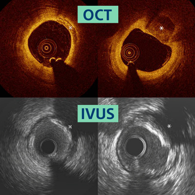
Two types of high definition intravascular imaging are commonly used in clinical practice: Intravascular Ultrasound (IVUS) and Optical Coherence Tomography (OCT).
IVUS employs sound waves to generate cross-sectional images of blood vessels, while OCT uses light waves to produce high-resolution images.
Both techniques offer unique advantages and play crucial roles in different clinical scenarios.
Importance of High Definition Intravascular Imaging
The importance of high-definition intravascular imaging lies in its ability to provide detailed information about vessel dimensions, plaque composition, and any abnormalities or obstructions. This knowledge aids medical professionals in diagnosing and treating conditions such as atherosclerosis, coronary artery disease, and structural heart defects.
By enabling precise visualization of the vessels, this technology assists in determining the most suitable treatment approach, reducing the risk of complications, and improving patient outcomes.
All About IVUS Guided Angioplasty
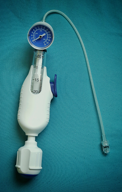
- IVUS-guided angioplasty is a procedure that combines intravascular ultrasound imaging with balloon angioplasty to treat coronary artery disease.
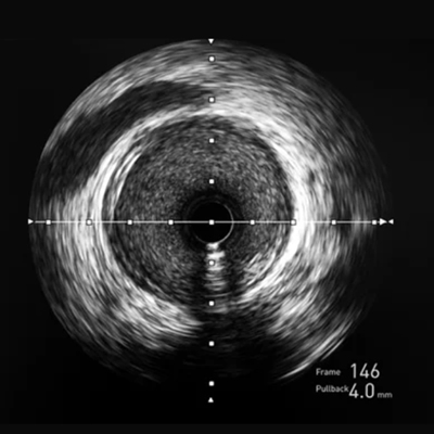
- A specially designed catheter with an ultrasound probe is inserted into the coronary arteries during the procedure. The probe emits sound waves and captures images of the vessel walls, providing real-time feedback to guide the placement of the angioplasty balloon.
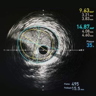
- The IVUS images obtained during the procedure help determine the optimal size of the angioplasty balloon and its exact positioning. This precise guidance ensures accurate stent placement and maximizes the effectiveness of the procedure.
- IVUS-guided angioplasty has been shown to improve outcomes by reducing the risk of complications such as stent malapposition, edge dissections, and residual stenosis.
By allowing the visualization of the vessel wall and the extent of plaque burden, IVUS helps identify high-risk lesions that may require additional treatment, such as plaque modification or stenting. This technique also aids in assessing stent expansion and apposition, ensuring optimal stent deployment and reducing the chances of restenosis.
Know About Coronary Intravascular Imaging
Coronary intravascular imaging refers to high-definition imaging techniques, such as IVUS and OCT, to visualize and assess coronary arteries. This imaging modality provides detailed information about the vessel’s structure, dimensions, and any abnormalities or plaque buildup.
Intravascular imaging of the coronary arteries has revolutionized the field of cardiology by offering precise and accurate assessments. It enables the evaluation of plaque characteristics, including composition, thickness, and vulnerability, helping identify high-risk lesions prone to rupture or causing blockages. This information assists in determining the appropriate treatment strategy, such as medical management, angioplasty, or stenting.
Moreover, coronary intravascular imaging is vital in guiding complex coronary interventions, such as chronic total occlusions and bifurcation lesions. It provides critical information regarding vessel anatomy, plaque distribution, and lesion severity, facilitating the selection of appropriate interventional techniques and devices.
Frequently Asked Questions
How safe is high-definition intravascular imaging?
High-definition intravascular imaging techniques, such as IVUS and OCT, are generally safe when performed by trained professionals. However, there may be a slight risk of complications associated with inserting catheters or using contrast agents.
Does high-definition intravascular imaging replace traditional angiography?
No, high-definition intravascular imaging does not replace traditional angiography. It complements angiography by providing additional information about vessel wall characteristics and plaque composition, improving diagnostic accuracy and treatment planning.
Can high-definition intravascular imaging detect all types of plaque?
High-definition intravascular imaging can detect various types of plaque, including calcified, fibrous, and lipid-rich plaques. However, individual plaques, such as microcalcifications or necrotic cores, may be challenging to visualize accurately.
How long does an IVUS-guided angioplasty procedure take?
The duration of an IVUS-guided angioplasty procedure varies depending on the complexity of the case and the operator’s expertise. On average, the procedure may take around 30 to 90 minutes.
Subscribe to Platinum For Heart Newsletter
Sign up now and get free access to our monthly newsletter on Heart health & More
Sources:
1. www.indianheartassociation.org
2. www.cardiosocietyindia.com
3. www.aiims.edu
4. www.medanta.org
Disclaimer: The information presented by Boston Scientific Corporation is for educational purposes only and does not recommend self-management of health issues. The information should not be treated as comprehensive and does not intend to provide diagnosis, treatment or any medical advice. Individual results may vary and hence, it is advisable to consult your doctor regarding any medical or health related diagnosis or treatment options.
IC-1660101AA-0823